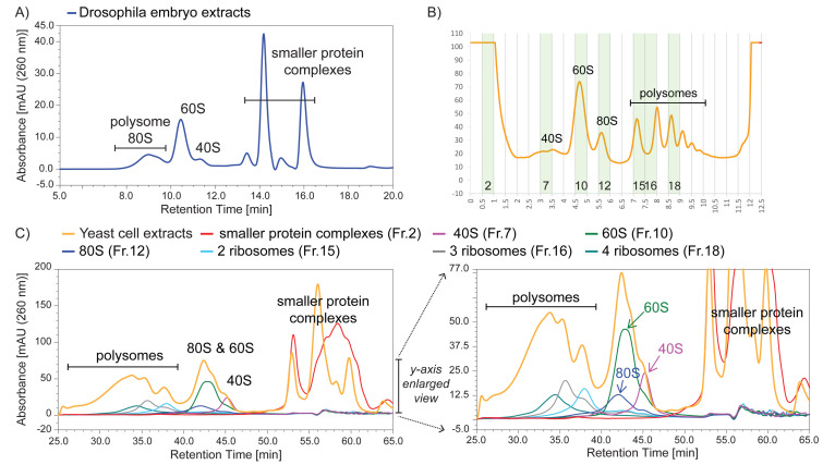
Figure 1. Ribo Mega-SEC profiles of Drosophila embryo and budding yeast cell extracts using a 1,000 Å pore size SEC column.
A) Drosophila embryo extracts were analyzed using a flow rate of 0.8 ml/min. B) Yeast cell extracts were analyzed by sucrose density gradient fractionation, and the highlighted fractions were collected for subsequent Ribo Mega-SEC analysis. C) Yeast cell extracts that were used for the sucrose density gradient analysis, or the fractions collected in Figure 2C, were injected onto a 1,000 Å pore size SEC column using a flow rate of 0.2 ml/min. The right panel shows a y-axis enlarged view of the left panel.

