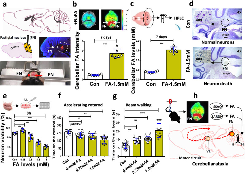
Fig. 6. Formaldehyde-infused into the fastigial nucleus promoted formaldehyde overload and motor deficits.
a Location, microinfusion, and identification of the bilateral fastigial nucleus injected with formaldehyde. FN fastigial nucleus (location identified by using red fluorescent probe-Dil to mark cell membrane and blue DAPI to stain nucleus). b Cerebellar formaldehyde quantified by an in-vitro small animal imaging system (n = 6). FA-1.5 mM: the group of mice microinfused with 1.5 mM formaldehyde; Con PBS-injected group of mice, FA formaldehyde. c Formaldehyde levels in the cerebellum detected by HPLC (n = 6). HPLC high-performance liquid chromatography. d Cellular morphology of the cerebellar neurons detected by using Nissl staining solutions (n = 3). e Cell toxicity of formaldehyde detected by using a CCK-8 kit (n = 6). f Motor behaviors assessed in FA-injected mice by the accelerating rotarod test (n = 10). g Motor functions in FA-injected mice evaluated by the beam walking test (n = 10). h The model of FN impairments results in cerebellar ataxia induced by HU-derived formaldehyde. VL ventrolateral nucleus of the thalamus. Error bars show the mean ± SEM; **p < 0.01; ***p < 0.001.

