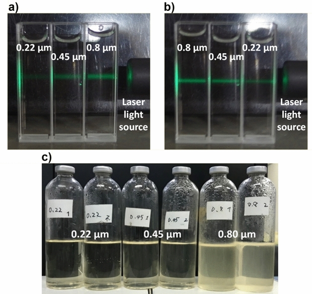
Figure 1.

Photo images of scattered light observed by irradiating green laser in the carbon dioxide nanobubbles medium which had been filtered with membrane filters and the culture media which were filtered with membrane filters and anaerobically cultured at 30 °C for 10 days. (a,b) The carbon dioxide nanobubbles medium was filtered with membrane filters whose pore size was 0.22 μm, 0.45 μm and 0.80 μm respectively. The green laser light irradiated the carbon dioxide nanobubbles medium from right to left, while the alignment of the samples was opposite each other in (a,b). It was clearly observed that the intensity of scattered light became lower as the pore size of filter which had been used for the filtration of the carbon dioxide nanobubbles medium became smaller, suggesting that more nanobubbles had been caught with the filter whose pore size was smaller. (c) Each two culture medium on the left, center and right had been filtered with 0.22 μm, 0.45 μm and 0.80 μm respectively. Every culture medium did not contain P. aeruginosa and the nanobubbles. The head space in the vial bottles were replaced with pure nitrogen. After 10 days cultivation, the culture media which had been filtered with 0.80 μm membrane filters were cloudy with bacteria whereas the culture media filtered with 0.22 μm and 0.45 μm were still remain clear, suggesting the indigenous bacteria had been completely sterilized with membrane filters whose pore size was less than 0.45 μm.
