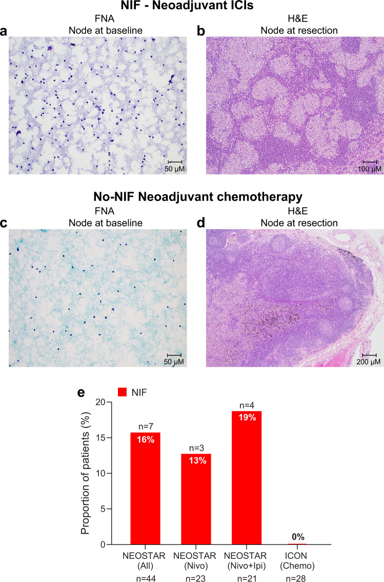
Fig. 2. Histopathological features of nodal specimens pre- and post-neoadjuvant therapy in NEOSTAR and ICON patients.
a Illustrative FNA image from preoperative mediastinal staging by EBUS in NEOSTAR NIF patient did not demonstrate granulomatous inflammation within examined nodes (station 4 L pictured; Papanicolaou, x20). b Resected nodal specimen in NEOSTAR NIF patient following ICIs demonstrating a diffuse non-caseating granulomatous inflammatory reaction (station 11 R pictured; hematoxylin and eosin, x10). c Illustrative FNA image from preoperative mediastinal staging by EBUS in ICON No-NIF patient did not demonstrate granulomatous inflammation within examined nodes (station 7 pictured; Papanicolaou, x20). d Resected nodal specimen following neoadjuvant chemotherapy in a patient with No-NIF from ICON cohort with the absence of diffuse non-caseating granulomatous inflammatory reaction (station 7 pictured; hematoxylin and eosin, x4). e Proportions of patients with NIF, characterized by abnormal nodes on imaging that are devoid of cancer and contain de novo non-caseating granulomas in NEOSTAR (n = 44) and ICON (n = 28) patient cohorts. The red bars depict the proportions of patients with NIF. Analyses related to the presented micrographs were conducted once. NIF, nodal immune flare; ICIs, immune checkpoint inhibitors; ICON, ImmunogenomiC prOfiling in NSCLC; EBUS, endobronchial ultrasound; FNA, fine needle aspiration. Source data for panel (e) are provided as a Source Data file.

