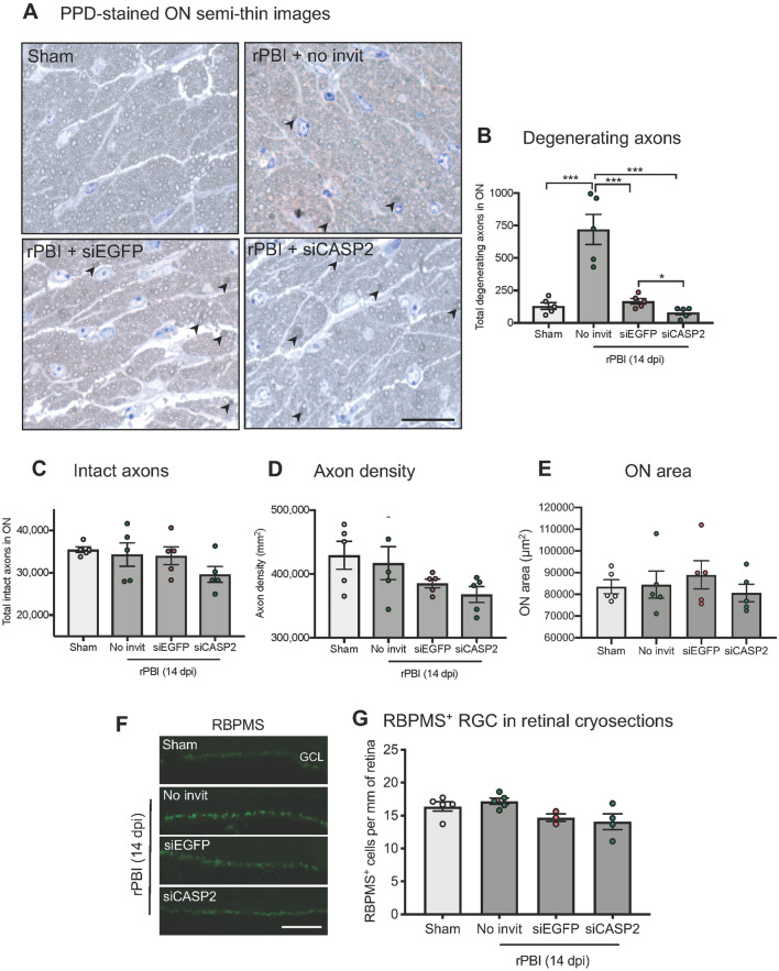
Figure 4.
Axonal counts in PPD-stained ON semi-thin sections and RBPMS+ RGC in retinal cryosections in the “pre-blast” injection study. (A) There were increased total degenerating axons in the ON after b-ITON compared to sham (P < 0.0001, ANOVA) but not in b-ITON eyes intravitreally injected with siCASP2 or siEGFP control (P < 0.001 for both siCASP2 and siCASP2 compared to b-ITON, post-hoc Tukey). The number of degenerating axons was lower in siCASP2 treated eyes compared to siEGFP controls, indicating a potential axon-protective effect of caspase-2 knockdown (P = 0.0170, post hoc Tukey). (B) There were no differences in intact axons between any groups (P = 0.214, ANOVA). (C) There was weak evidence of differences in ON axon density between sham and b-ITON groups (P = 0.1099, ANOVA). (D) Representative PPD and toluidine blue-stained resin semi-thin ON sections, with degenerating axons (arrowheads). (E) There were no differences in ON area between groups (P = 0.7244, ANOVA). (F) Representative IHC images showing RBPMS+ RGC in the GCL on retinal cryosections. (G) There was weak evidence of a difference in the number of RBPMS+ RGC after b-ITON with “pre-blast” intravitreal injections (P = 0.1364, ANOVA). Scale bar in (D) = 20 μm and (E) = 100 μm. Error bars represent mean ± SEM.

