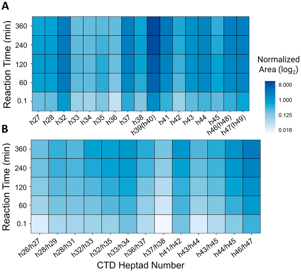
Figure 6.

Heat maps illustrating the time-dependent variations in Ser5 phosphorylation by CDK7 of human distal CTD. (A) Mono-phosphorylated heptads. (B) Bis-phosphorylated heptads with pairs of Ser5 positions separated by a slash mark. h39(h40), h46(h48), and h47(h49) refer to the YSPTSPTYSPTSPK peptides, which are repeated twice and thus correspond to phosphorylation of sites on heptads 39 or 40, 46 or 48, and 47 or 49. The y-axis is normalized using log2 area and shows phosphorylation levels at each Ser5 site localization stacked for each time point.
