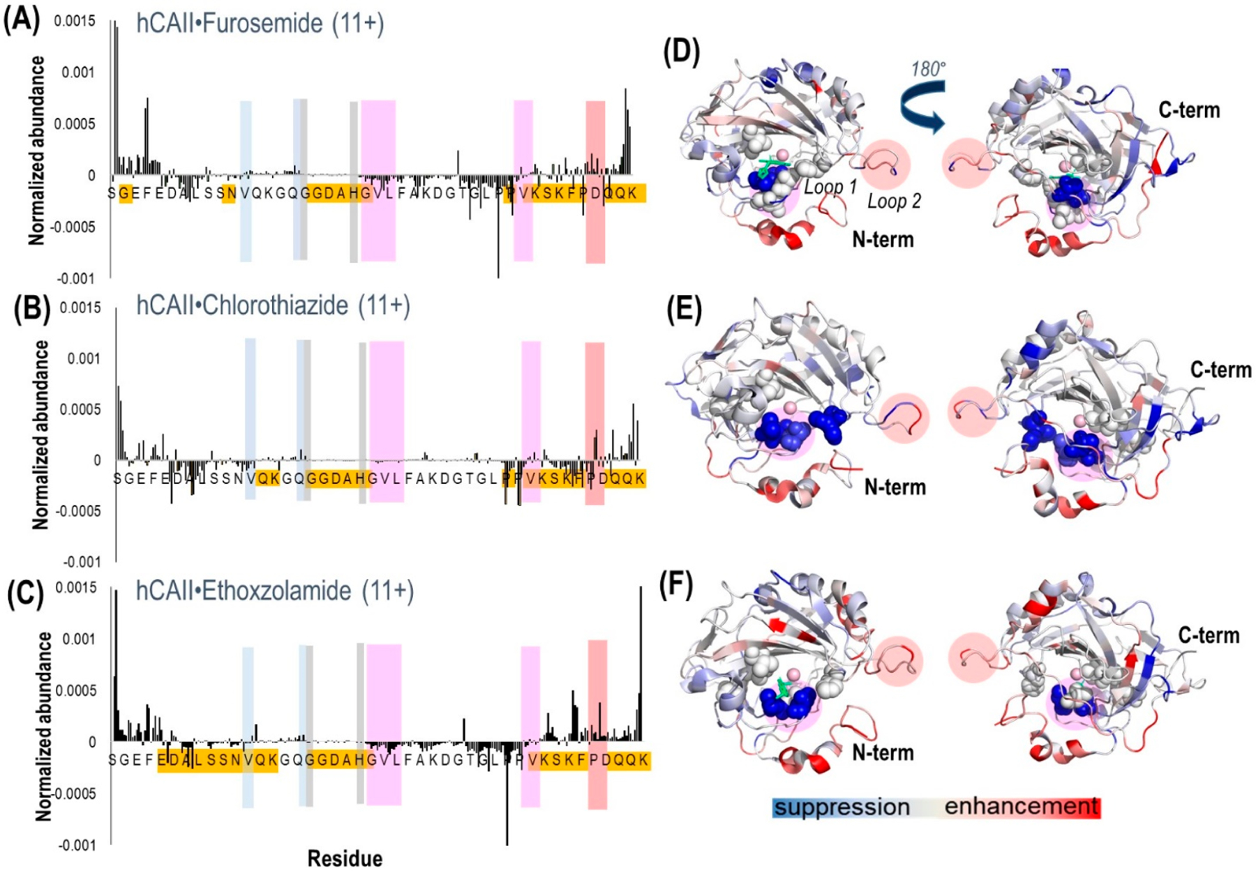
Figure 5.

Plots of the differences in abundances of fragment ions (both apo and holo ions) obtained from hCAII complexed with (A) furosemide, (B) chlorothiazide, and (C) ethoxzolamide. Highlighted are the hydrophobic pocket/loop 1 (pink), the hydrophilic pocket (light blue), the residues that coordinate zinc (gray), and loop 2 (peach). Highlighted in gold are the residues where the most meaningful differences in backbone fragmentation occurred. The crystal structure of hCAII shows the suppression (blue-shaded residues) or enhancement (red-shaded residues) of backbone cleavages upon UVPD for the complexes containing (D) furosemide (PDB 1Z9Y), (E) chlorothiazide (PDB 2ILI), and (F) ethoxzolamide (PDB 3CAJ). For the structures, the sulfonamide ligand is depicted in green. There is no reported crystal structure of hCAII–chlorothiazide, so the structure is shown for hCAII. Regions shaded in a pink circle (residues 197–206) indicate loop 1, and those shaded in a peach circle (residues 230–240) indicate loop 2. The residues corresponding to the hydrophobic pocket (V121, V143, L198, T199, H200, W209) are represented as spheres.
