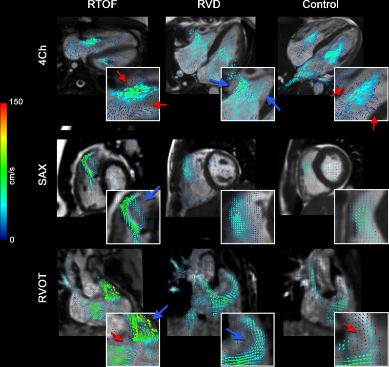
Fig. 4.
Qualitative intracardiac flow profile via velocity vector fields overlaid onto cine imaging. Blue arrows are pointed to larger vorticity magnitude, red arrows pointed to smaller vorticity magnitude. In rTOF patients, there is a dominant vortex generated from pulmonary insufficiency, best visualized in short axis (SAx) and modified right ventricular outflow tract (RVOT) view. This vortex is separate from the smaller vortices generated by the tricuspid inflow, best visualized in the 4-chamber (4Ch) view. For right ventricular dilation (RVD patients), a larger vortex is generated from tricuspid inflow and propagates into the RVOT, but the overall flow topology is unchanged when compared to controls

