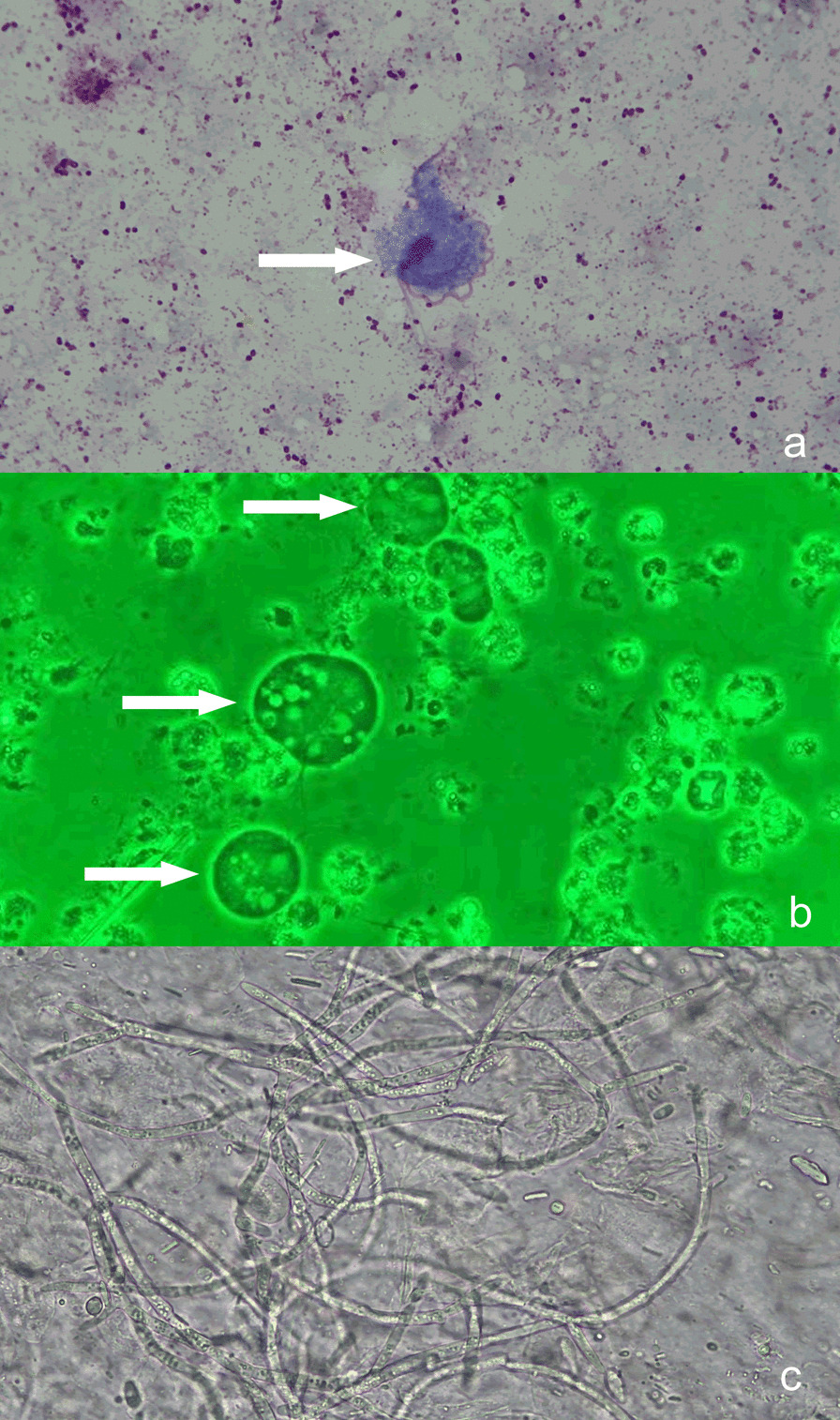
Fig. 2.

a Bright-field microscopy with Wright's-Giemsa staining (× 1000). b Phase-contrast microscopy of a sample taken during exercise (wet smear, × 1000). As indicated by the arrows, the 4 characteristic free anterior flagella, nucleus, and axial column of the T. tenax trophozoites were well stained. The undulating membrane is shorter than the long axis of the trophozoite, and accounts for about half of the whole trophozoite body. The slender axial column runs through the trophozoite and extends out of the body from the back, and the axostyle is relatively thick. The nucleus is located in the anterior part of the trophozoite, and has an oval shape with many chromatin granules. c Sputum smear, showing a large number of hyphae from G. capitatum (× 1000)
