
Fig. 1. Rat NPCs extend greater numbers of axons into spinal cord white matter than gray matter.
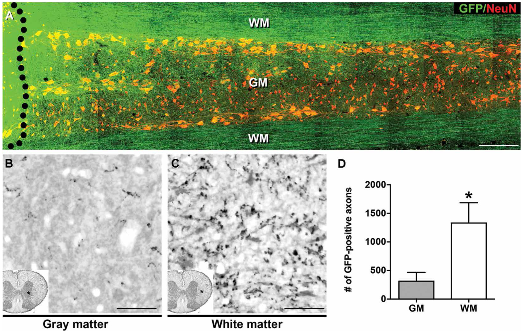
(A) GFP-expressing E14 rat spinal cord–derived NPCs were grafted into sites of C5 spinal cord hemisection lesions (black dotted line indicates graft boundary) 2 weeks after injury. One month after grafting, horizontal sections of spinal cord were immunolabeled for GFP (green) and the neuronal marker NeuN (red). Immunolabeling indicated that rat NPCs extended their axons through spinal cord host white matter (WM) and gray matter (GM). Greater numbers of axons appeared to extend through WM than GM. The rostral section of spinal cord is on the left, and the caudal section is on the right. (B and C) GFP immunolabeling of rat NPC graft–derived axons in transverse sections of rat spinal cord demonstrated larger numbers of axons extending into caudal host spinal cord white matter, three spinal segments below the graft site. (D) Quantification of data in (B) and (C). Mean ± SEM (*P < 0.05, paired, two-tailed t test; n = 4 rats). Scale bars, 500 μm (A), 25 μm (B and C).