FIGURE 11.

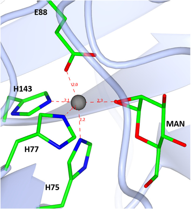
A diagram showing the D-mannose sugar bound in the active site of TsLI with the manganese ion (shown in red) co-ordinated to the three histidines H75, H77 and H143, one glutamic acid E88, and the manganese ion.

A diagram showing the D-mannose sugar bound in the active site of TsLI with the manganese ion (shown in red) co-ordinated to the three histidines H75, H77 and H143, one glutamic acid E88, and the manganese ion.