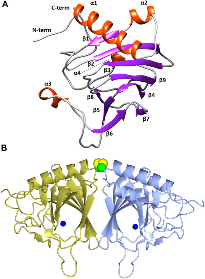
FIGURE 5.

(A) A cartoon representation of the TsLI monomer with the secondary structure elements labelled. Figure was prepared with UCSF Chimera (Pettersen et al., 2004). (B) A cartoon representation of the cupin fold architecture of the TsLI dimer. The bound manganese ions are shown as blue spheres and the disulfide bond between the cysteines C22 of each monomer are shown as a space filling model. Figures 5B, 8–12 were prepared with CCP4mg (McNicholas et al., 2011).
