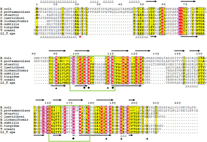
FIGURE 6.
Alignment of the amino acid sequences of various LIs. Residues related to substrate binding sites (black circle), and those involved in both the metal coordination and substrate binding sites (black triangle), according to the ligand bound structures of E. coli LI bound to D-fructose (PDB 3KMH) and B. subtilis LI (PDB 2Y0O). Identical residues are in red while similar residues are in yellow, the green box highlights the cupin family motif 1 and 2. The red star indicates the cysteine 22 which is responsible of the disulfide bond connecting the dimer of the TsLI. The secondary structure of the D-LI from E. coli and TsLI are represented at the top and bottom of the alignment respectively with spring and arrow indicating α helices and β sheets. The alignment was carried out using ESPript 3.0 (http://espript.ibcp.fr/ESPript/ESPript/) (Robert and Gouet, 2014).

