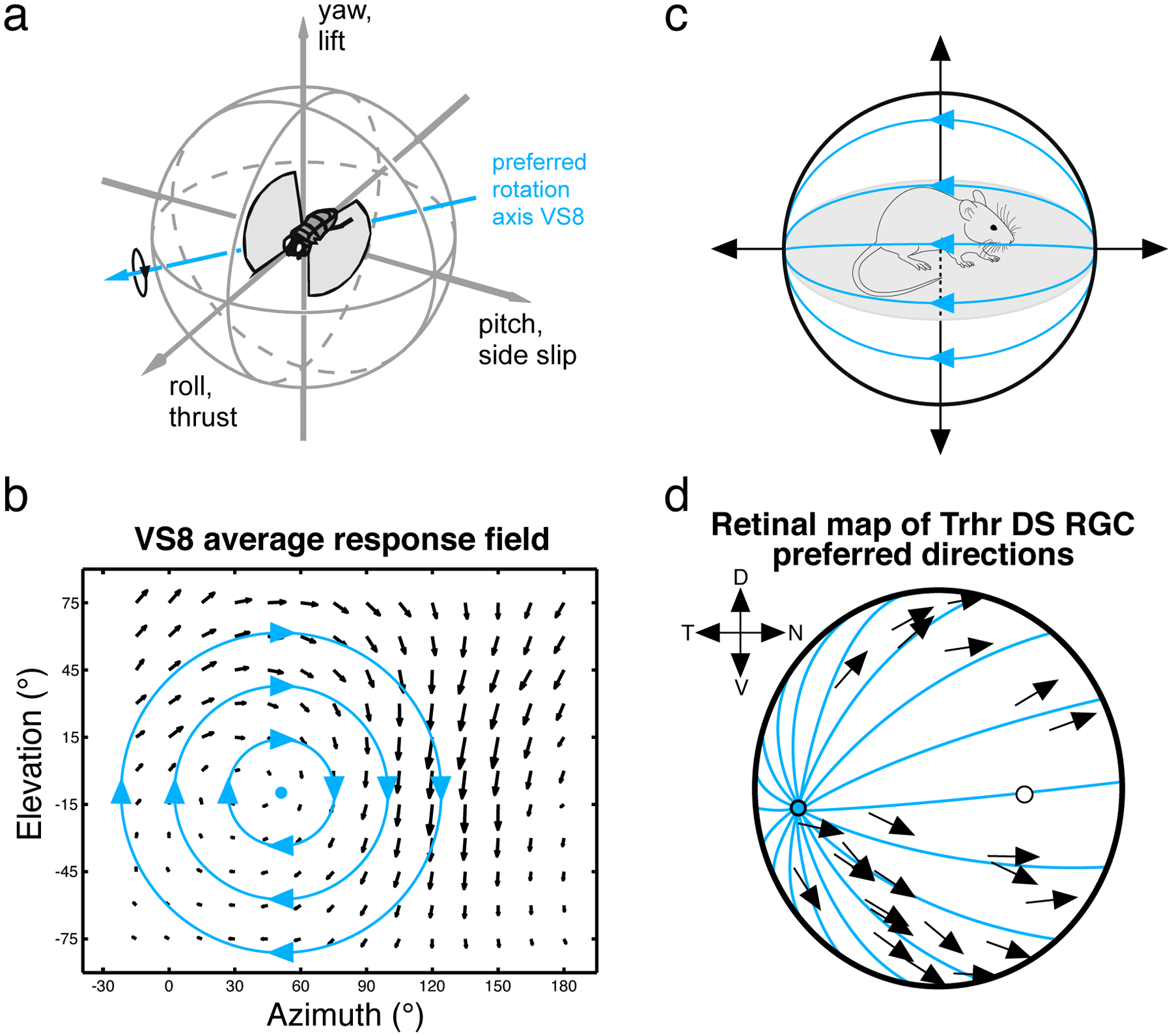
Figure 4: Motion sensitive neurons encode self-movement across the animal kingdom.

(a) Schematic showing a fly in flight. (b) Local motion receptive field of the VS8 neuron in the blowfly Calliphora. The direction of each arrow indicates the local preferred direction, and the length of each arrow indicates the cell’s motion sensitivity. This local motion receptive field corresponds to the optic flow pattern that would result from a rotation of the animal. The rotation axis around which the fly would need to turn to maximally activate this neuron is indicated in (a). Data & schematic provided by Holger Krapp. (c) Schematic showing a mouse ambulating in a forward direction. The resulting visual input is an optic flow pattern emanating from a singularity directly ahead of the animal (blue lines). (d) Direction preferences of a population of DS RGCs in mouse retina are overlaid on the retinal surface. Forward motion optic flow moves outward from a point in the retina (blue lines). The direction preferences of this cell type roughly align with the optic flow lines that result from forward motion. Other DS RGC types similarly respond to optic flow resulting from other directions of motion of the animal. Data redrawn from (Sabbah et al., 2017).
