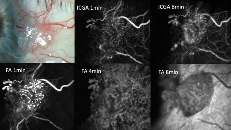
Figure 1.
Slit lamp photograph, ICGA and fluorescein angiograms of a case of conjunctival squamous cell in situ carcinoma. Pronounced focal early indocyanine green dye leakage is visible in the upper middle image. Both focal and diffuse indocyanine green leakage are visible in the upper right image. The lower images show diffuse dye leakage in all phases of fluorescein angiography (FA). Note that fluorescein leakage occurs in both tumour and healthy surrounding conjunctiva, while indocyanine green leakage is exclusively confined to the lesion. ICGA. indocyanine green angiography.

