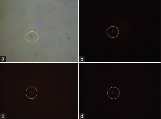
Figure 2.

The illustration of circulating tumor cells isolated from whole blood sample of a breast cancer patient. Panel (a) represent light microscopy image of isolated circulating tumor cell. Captured cells were stained with Hoechst®33342 dye (b), with an anti-cytokeratin FITC-conjugated antibody (c), and with an unconjugated β4 integrin antibody and PE anti-mouse IgG1 Antibody (d)
