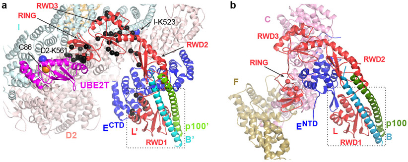
Figure 2 ∣. FANCL at the active and inactive sides of Core-UBE2T-ID-DNA.
a-b, Close-up views of the active (a) and inactive (b) sides of the Core-UBE2T-ID-DNA complex showing FANCC-FANCE-FANCF sequesters FANCL’ residues required for ID and FANCECTD binding (only subunits or portions thereof near FANCL’ are shown). Active-side FANCL’ residues marked with black Cα spheres correspond to residues sequestered within 3.8 Å of FANCC- FANCENTD-FANCF at the inactive side. Dotted box indicates the portions of FANCL-FANCB-FAAP100 aligned to equivalently orient the views of the two sides. In a, FANCI Lys523 and FANCD2 Lys561 are marked by large blue spheres, and UBE2T Cys86 by an orange sphere.

