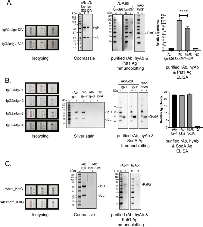
Fig 4. IgV sequence validation of PstS1NRC-2410, SodANRC-13810, and KatGNRC-49680 hybridomas.
The correct IgV sequences of PstS1NRC-2410 (A), SodANRC-13810 (B) and KatGNRC-49680 (C) were identified from multiple IgVH and IgVL sequences (S3 and S4 Tables). Each heavy and light chain combination of the isotype-switched IgG2a and Igκ constructs was separately co-expressed in 293 F cells by transient transfection. The presence of an assembled, secreted rAb can be detected in the culture supernatant on Day-6 post-transfection using a mouse Ig Isotype kit (Isotyping label). Some combinations yield an undetectable Ig in the cellular supernatant. The cellular supernatant of each combination was collected and purified by protein A column chromatography. The Ig-equivalent elution fractions were pooled and concentrated. Coomassie or silver staining shows the purity of the rAbs. The Ag-Ab binding potential of each IgVH/IgVL pair was compared to that of the corresponding hyAb. Equivalent amounts of either the rAb or hyAb were kept in immunoblot and ELISA assays to ensure equitable comparison. Immunoblot assays detected recognition of their cognate purified antigen (Immunoblotting). The antigen-binding potency was quantified by ELISA using purified antigens. Relative Ig affinity was determined by ELISA endpoint titers normalized against that of an ELISA buffer control (negative control, NC). Data shown here are from two independent experiments performed in triplicate. P-value<0.001 indicated by the asterisks.

