
Figure 3.
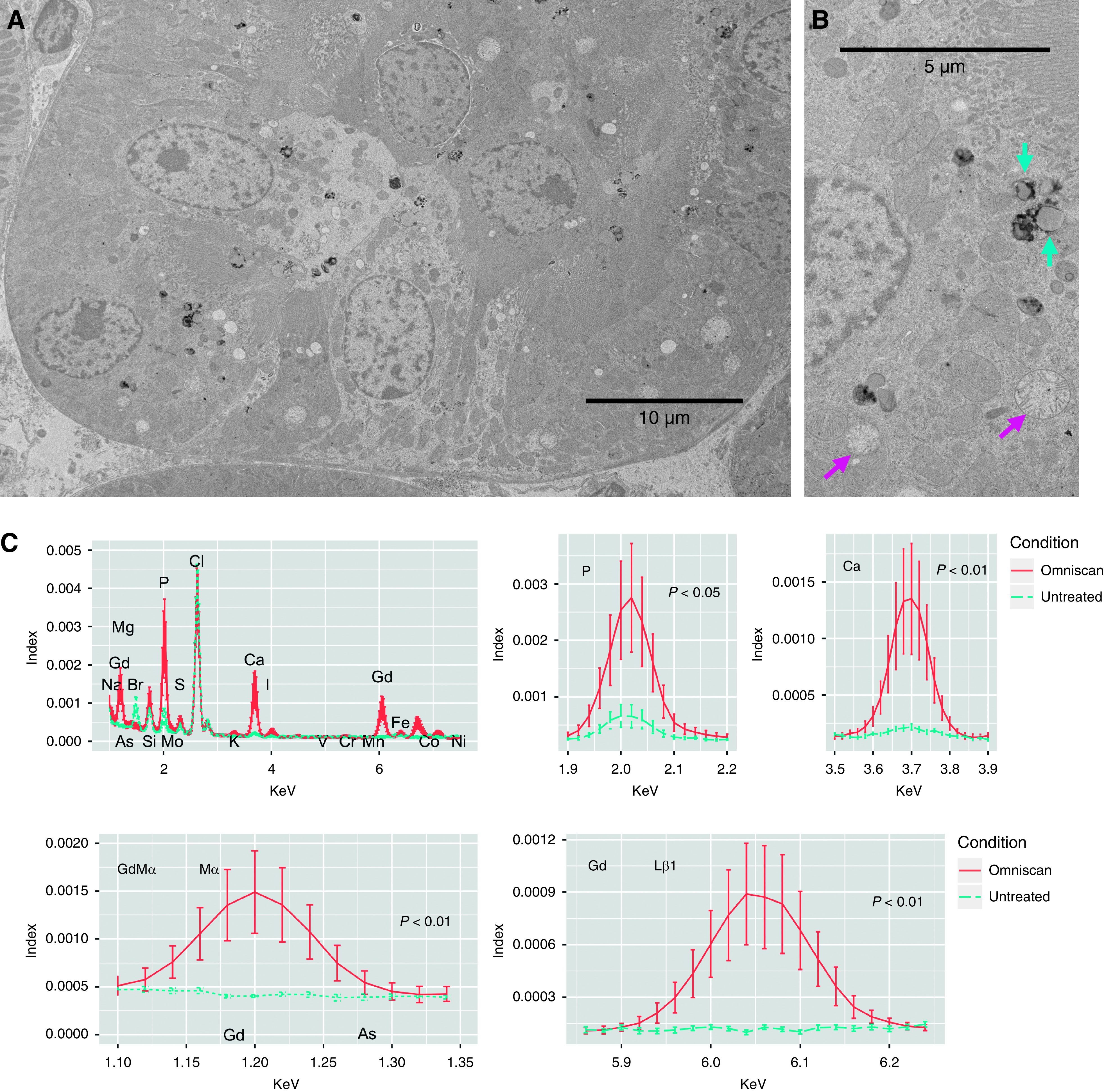
Gadolinium-based contrast agents ravage the renal proximal tubule. (A) Invariably, treatment with gadolinium-based contrast agent leads to a peppering of the electron-dense nanostructures in lipid-laden vacuoles of proximal tubular cells. The low-magnification transmission electron photomicrograph demonstrates active tubular damage. (B) The magnified view highlights the lipid-laden vacuoles (cyan arrows) and widespread mitochondrial death (magenta arrows). (C) The electron-dense nanostructure precipitates contain gadolinium, phosphorus, and calcium. Sections (200-µm thick, epoxy embedded) were analyzed by scanning transmission electron microscopy equipped with energy-dispersive x-ray spectroscopy. Spectra for the electron-dense precipitates (n=13) and renal cortex from an untreated animal (n=6 measurements) were normalized. Areas under the curves were compared using William Sealy Gosset two-tailed t tests. JEOL 2010F FEGSTEM operating at 200 kV with Oxford Analytical Aztec energy-dispersive x-ray spectroscopy system equipped with an XMax 80N 80-mm2 silicon drift detector.