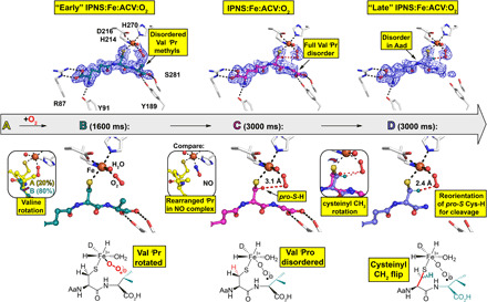
Fig. 3. tr-SFX reveals mode of O2 binding to IPNS:Fe:ACV.

ACV-derived conformations (A to D) were fit and refined to the 1600-ms (ACV conformation A and B) and 3000-ms (ACV conformation C and D) tr-SFX datasets (PDB: 6ZAI and 6ZAJ). 2mFo-DFc omit maps (1.0 σ contour level, 1.53 and 1.55 Å resolution, respectively) are shown. Conf. A: The same as the anaerobic ACV complex (Fig. 1). Conf. B: The major 1600-ms IPNS:Fe:ACV + O2 conformation (B, 80%) refined with the ACV Val isopropyl methyls deleted (teal). Left inset: Overlay of IPNS:Fe:ACV (A, 20%, details in Fig. 1B) and IPNS:Fe:ACV:O2 (B, 80%) 1600-ms models. Conf. C: (3000 ms), where all Val isopropyl atoms are disordered. Central inset: The anaerobic IPNS:Fe:ACV:NO complex (PDB: 6ZAN, cryo MX, 1.39-Å resolution). Conf. D: New proposed conformation in the 3000-ms dataset with the Cys side-chain rotated. Right inset: Superimposition of confs. C and D, showing how the ACV cysteinyl methylene rotation positions the pro-S CCys,β─H bond close to the distal O of the Fe-O2 for stereospecific C─H cleavage (fig. S6).
