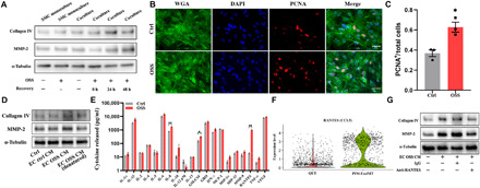
Fig. 4. OSS-induced EC phenotypic changes cause proliferation and ECM protein remodeling in SMCs via RANTES-mediated paracrine mechanism.

(A) ECs and SMCs were cocultured on the microchip, and the chips were incubated for another 0 to 48 hours after ECs were exposed to OSS stimulation for 24 hours. Representative immunoblotting showing increased collagen IV and MMP-2 levels in SMCs. (B) Immunofluorescence images displaying increased expression of PCNA in SMCs. (C) PCNA+ cells were counted three times in at least four high-power fields by two evaluators. Scale bars, 50 μm. Data are expressed as means ± SEM of three independent experiments. *P < 0.05. (D) SMCs were treated with different CM as indicated for 48 hours, and the protein levels of collagen IV and MMP-2 were measured by Western blot analysis. (E) Multiplex ELISA for EC pro-inflammatory cytokine and chemokine expression with or without OSS treatment. Data are expressed as means ± SEM of four independent experiments. *P < 0.05 and **P < 0.01. (F) Violin plots showing relative expression of RANTES (CCL5) in QET and pro-inflammatory EndMT clusters. (G) SMCs were treated for 48 hours with EC OSS CM preincubated with RANTES neutralizing antibody (10 μg/ml) or nonimmune mouse immunoglobulin G (IgG; 10 μg/ml) for 1 hour, and the protein levels of collagen IV and MMP-2 were measured by Western blot analysis. Densitometric data of immunoblots are presented in fig. S6.
