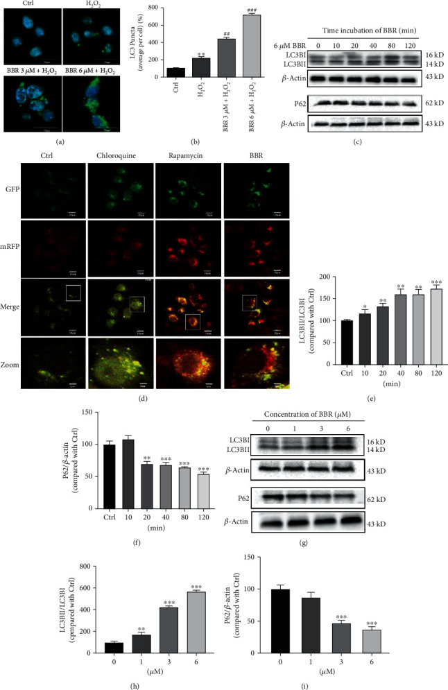
Figure 2.

BBR stimulates autophagy in D407 cells. (a) Confocal microscopy examination of LC3B expression. (b) Quantification of LC3B puncta. (c) D407 cells were treated with 6 μM BBR for different time periods as indicated, and LC3B, P62, and β-actin were detected by western blotting with specific antibodies. (d) Autophagic flux was detected by LC3 double fluorescent lentivirus autophagy flow detection system; a tandem GFP-RFP-LC3 fusion protein was treated with chloroquine (CQ, 60 μM), rapamycin (1 μM), and berberine (BBR, 6 μM) for 2 h. Confocal microscopy was used to examine the autophagic flux (scale bar = 10 μm). (e, f) Quantification of the representative protein bands from western blotting. (g) D407 cells were treated with various concentrations of BBR for 2 h, and the expression of LC3B, P62, and β-actin was detected by western blotting with specific antibodies. (h, i) Quantification of the representative protein bands from western blotting. The assay was repeated for at least 3 times. ∗p < 0.05, ∗∗p < 0.01, and ∗∗∗p < 0.001 versus the control group; ##p < 0.01, ###p < 0.001 versus the H2O2-treated group were considered significantly different.
