Figure 5.

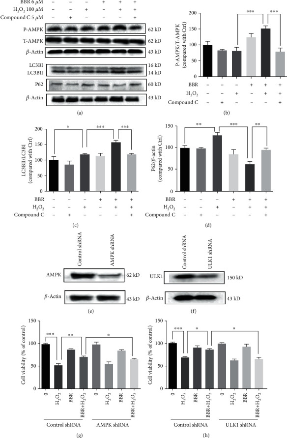
AMPK specific inhibitor compound C blocked the autophagy-stimulating and protective effect of BBR. (a) D407 cells were pretreated with 5 μM compound C for 30 min and 6 μM BBR for 2 h and then incubated with or without H2O2 for further 2 h. The expression of phosphorylated AMPK, total AMPK, LC3B, P62, and β-actin was detected by western blotting. (b–d) Quantification of the representative protein bands from western blotting. (e) Cells were transfected with AMPK shRNA for 48 h, and the expression of AMPK was detected by western blotting. (f) Cells were transfected with AMPK shRNA, treated with 6 μM BBR or 0.1% DMSO (vehicle control) for 2 h, and then incubated with or without 100 μM H2O2 for 24 h. Cell viability was measured by MTT assay. (g) Cells were transfected with ULK1 shRNA for 48 h, and the expression of ULK1 and β-actin was detected by western blotting. (h) Cells were transfected with ULK1 shRNA, treated with 6 μM BBR or 0.1% DMSO (vehicle control) for 2 h, and then incubated with or without 100 μM H2O2 for 24 h. Cell viability was measured by MTT assay. The assay was repeated for at least 3 times. ∗p < 0.05, ∗∗p < 0.01, and ∗∗∗p < 0.001 were considered significantly different.
