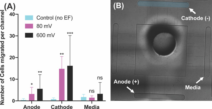
Figure 3.
Neutrophil-like cells electrotaxis under the effect of the DC electric field. (A) Quantification of the number of neutrophils migrated per channel toward the cathode. The result shows a significant increase (65%-80%) in migration by applying different DC electric field strength (n=4, p-value<0.005). Quantification of the number of neutrophils migrated per channel toward the anode. The result indicates a significantly low, less than ~5 cells per channel, directional movement of neutrophils toward the anode. Quantification of the number of neutrophils migrated per channel toward the complete media. The result shows a significant low, less than ~3 cells per channel, migration toward the complete media due to no electrical or chemical signals. (B) Nikon TiE microscope 10X image of the microfluidic device and experiment setup of electrotaxis. *P ≤ 0.05; **P ≤ 0.01; ***P ≤ 0.001; ns, not statistically significant.

