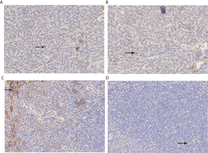
Figure 1.
PDL1 immunohistochemical analysis. (A, B) Immunohistochemical analysis of PDL1. The percentage of positive-stained cells was 1% (X200); (C) Positive control image of immunohistochemical analysis of PDL1 (X200). (D) Negative control imaging of immunohistochemical analysis of PDL1 (X200). PDL1, programmed cell death ligand 1.

