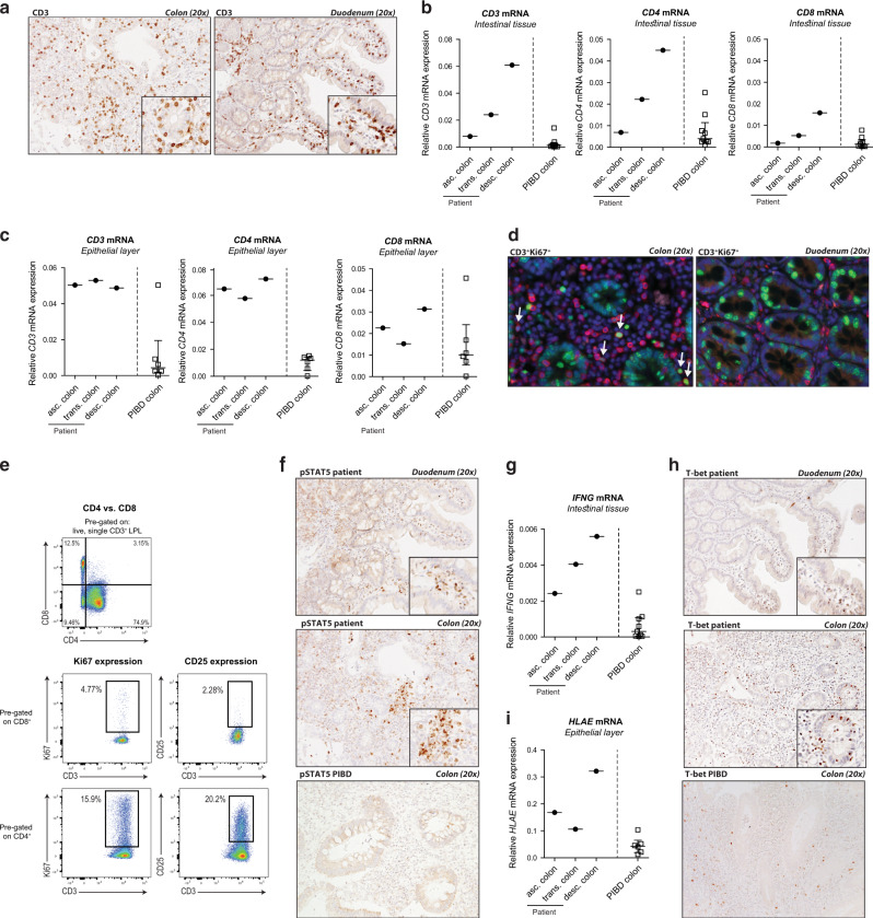
Fig. 3. Increased proliferation and activation of colonic CD4+ IEL and LPL in the patient with IL2RA duplication compared to treatment-resistant pediatric-onset IBD.
a, f, h Representative immunohistochemical staining for CD3, pSTAT5, and Tbet in paraffin-embedded resected inflamed colonic tissue (visit S2) and paraffin-embedded tissue of the unaffected duodenum from the patient at time of diagnosis (visit S0) and PIBD control. b CD3, CD4, and CD8 mRNA and g IFNG mRNA expression in total resected colonic tissue of the patient and PIBD controls. c CD3, CD4, and CD8 mRNA and (i) HLAE mRNA expression in the epithelial layer isolated from resected colonic tissue of the patient and PIBD controls. d Representative immunofluorescent double staining of paraffin-embedded resected colonic tissue of the patient. Green = Ki67, red = CD3, blue = 4′,6-diamidino-2-phenylindole (DAPI) nuclear staining. e LPL were isolated from the inflamed colonic tissue of the patient. Frequencies of CD4+ and CD8+ cells in live CD3+ LPL, and Ki67 and CD25 expression by CD4+ and CD8+ LPL were analyzed by flow cytometry.

