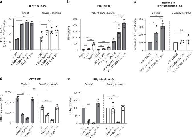
Fig. 4. Increased IL-2 signaling enhances IFNγ production and is reversed by JAK1/3 inhibition.
a–c PBMCs of the patient and healthy adult controls were stimulated with anti-CD3 (500 ng/ml) or anti-CD3/anti-CD28 beads (bead-to-cell ratio 1:2) in the absence or presence of IL-2 (1, 50, or 100 IU/ml) for 48 h. a Percentage of IFNγ-expressing CD4+ T cells were analyzed by flow cytometry. b IFNγ secretion by patient cells was analyzed using ELISA. c IFNγ responses of patient and healthy adult donors are shown. The relative increase in IFNγ secretion between anti-CD3/CD28 and cultures with IL-2 is shown (considering the percentage of cytokine secretion upon anti-CD3/CD28 stimulation as 100%). d, e PBMCs of the patient and healthy adult controls were stimulated with anti-CD3/anti-CD28 beads (bead-to-cell ratio 1:2) in the absence or presence of tofacitinib (200 or 1000 nM) for 48 h. d CD25 expression on CD4+ T cells was analyzed by flow cytometry. e Supernatants were assayed for IFNγ using an ELISA. The relative difference in IFNγ secretion between anti-CD3/CD28 and cultures with tofacitinib is shown (considering the percentage of cytokine secretion upon anti-CD3/CD28 stimulation as 100%). Data are mean ± SEM (adult healthy controls, n = 4); n.s., not significant, *p < 0.05, **p < 0.01, ***p < 0.001 using one-way ANOVA followed by the Bonferroni’s Multiple Comparison Test.

