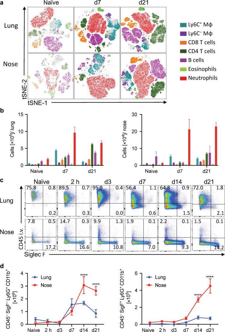
Fig. 1. Siglec-F+ neutrophils are recruited to the nasal tissue during B. pertussis infection.
C57BL/6 mice were aerosol infected with B. pertussis. At different time points, mice were injected i.v. with fluorochrome-labeled CD45 antibody and euthanized 10 min later. Cell suspensions were prepared from lung and nasal tissue and immune cells were analyzed by flow cytometry. a Representative tSNE plots for cells from lung and nasal tissue in naive mice and 7 or 21 days post infection. Neutrophils (Ly6G+Ly6C+), eosinophils (Siglec-F+ CD11c−), B cells (B220+MHCII+), CD4 T cells (CD3+CD4+), CD8 T cells (CD3+CD8+), Ly6C− macrophages (F4/80+CD11b+Ly6C−), Ly6C+ macrophages (F4/80+CD11b+Ly6C+). b Absolute cell counts for immune cell populations in lung and nose. c Cells were pre-gated on total neutrophils (Ly6G+CD11b+), dot plots show the intravital CD45 stain on the y-axis and Siglec-F on the x-axis. d Absolute cell numbers of tissue-resident Siglec-F− neutrophils (CD45 i.v.− Siglec-F−Ly6G+CD11b+) and Siglec-F+ neutrophils in lung and nose. n = 4/group, mean ± SEM, statistical analysis: Two-way ANOVA followed by Sidak’s post-test, significances are indicated in comparison to naive mice of the same population, ****p < 0.0001.

