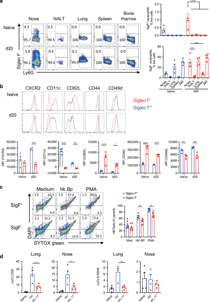
Fig. 10. Siglec-F+ neutrophils have an activated phenotype and high NET activity.
C57BL/6 mice were aerosol infected with B. pertussis and euthanized on 20 d post challenge. Cell suspensions were prepared from several tissues and immune cells were analyzed by flow cytometry. a Representative dot plots showing Siglec-F+ expression on CD11b+Ly6G+ pre-gated cells and percentages of Siglec-F+ and Siglec-F− neutrophils in naive mice and on day 20 post challenge in nasal tissue, NALT, lung, spleen, and bone marrow. b Histograms and MFIs showing the expression of the surface markers CXCR2, CD11c, CD62L, CD44, and CD49d on Siglec-F+ and Siglec-F− neutrophils in nasal tissue of naive mice and mice 20 d post challenge with B. pertussis. c Single cell suspension was prepared from nasal tissue of B. pertussis infected mice up to 1-month post challenge, was stimulated with heat-killed B. pertussis or PMA and then stained with the intracellular DNA dye (DAPI) and the cell-impermeant DNA dye (SYTOXgreen). Percentage of cells undergoing NETosis (double positive for DAPI and SYTOXgreen). d mRNA was prepared on day 6 post re-challenge from lungs and nasal tissue of convalescent mice treated with anti-IL-17A or isotype. Gene expression of Lcn2 and S100a8 was determined by RT-PCR and normalized against 18S RNA. Statistical analysis: a Two-way ANOVA followed by Tukey’s post-test, b, c Two-way ANOVA followed by Sidak’s post-test, d One-way ANOVA followed by Tukey’s post-test, *p < 0.05, **p < 0.01, ***p < 0.001; a, b, d n = 4/ group, c n = 7/group, pooled from two independent experiments, mean ± SEM.

