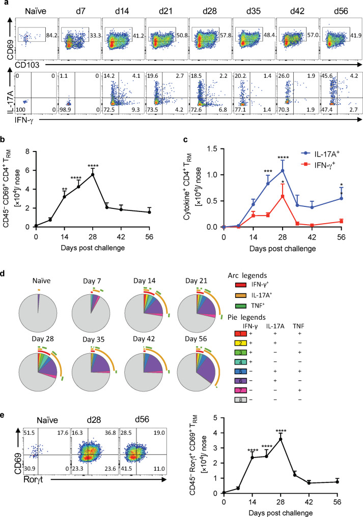
Fig. 2. IL-17A-secreting CD4 TRM cells accumulate in the nasal tissue during B. pertussis infection.
C57BL/6 mice were aerosol infected with B. pertussis. At different time points, mice were injected i.v. with fluorochrome-labeled CD45 antibody and sacrificed 10 min later. Cell suspensions were prepared from nasal tissue and cells were stimulated with sBP and anti-CD49d/CD28 and analyzed by flow cytometry. a CD69 and CD103 expression on tissue-resident CD44+ CD4+ T cells and IL-17A and IFN-γ-production in the CD69+ sub-population. b Total number of CD4 TRM (CD45 i.v.− CD4+ CD44+ CD69+) in nasal tissue during B. pertussis infection. c Total number of B. pertussis-specific IL-17A- or IFN-γ-producing CD4 TRM cells. d SPICE analysis of cytokine production in sBP-stimulated CD4 TRM. e Analysis of RORγT expression in CD4 TRM cells in naive mice and 28 and 56 days post infection. Absolute numbers of tissue-resident RORγt+ CD69+ CD4 T cells. Statistical analysis: b One-way ANOVA followed by Dunnett’s post-test, significances are indicated in comparison to naive mice; c Two-way ANOVA followed by Sidak’s post-test, significances are indicated in comparison to naive mice of the same population; *p < 0.05, **p < 0.01, ***p < 0.001, ****p < 0.0001, n = 4/group, mean ± SEM.

