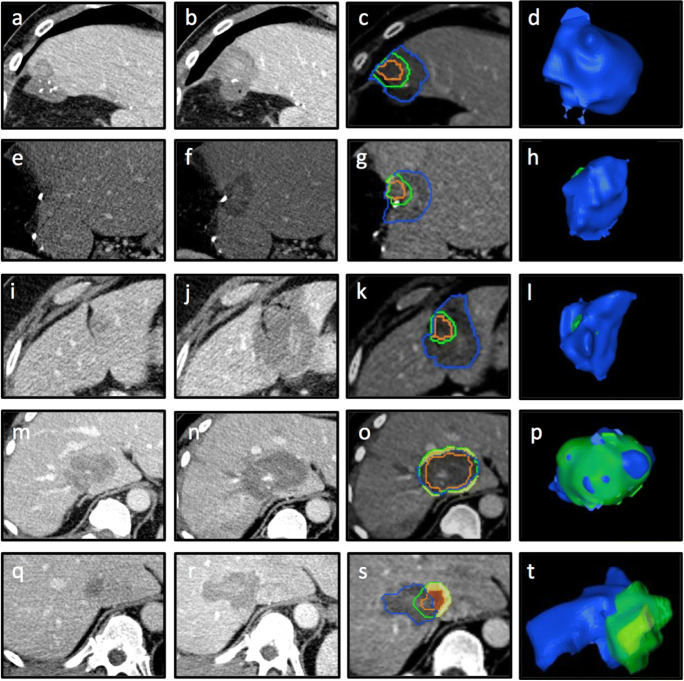
Fig. 3.
Examples of periablational 3D safety margin (SM) assessment with Ablation-fitTM. Complete ablation with 100% 3D SM (a–d), 95–100% 3D SM (e–h), 90–95% 3D SM (i–l), < 90% 3D SM (m–p), and incomplete ablation with residual tumor (q–t). a, e, i, m Pre-interventional CT-scan. b, f, j, n Post-interventional CT-scan. c, g, k, o Non-rigid registration of pre- and post-interventional CT scans with assessment of 3D SM of 5 mm: tumor (orange line), 5-mm safety margin (green line), necrosis zone (blue line), unablated safety margin (yellow area); unablated tumor (red area). d, h, l, p 3D reconstruction of necrosis zone (blue), unablated 3D safety margin (green), and unablated residual tumor (yellow)

