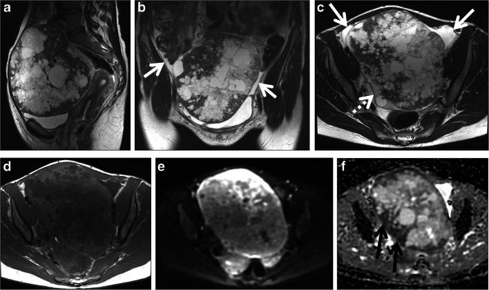
Fig. 3.
A 37-year-old woman with a pelvic mass. CA125 was 34 kU/L. Sagittal (a), coronal (b), axial T2-weighted (c) and axial T1-weighted (d) images show a large pelvic mass with solid and cystic areas. Small-volume ascites is seen around the mass (white arrows in b and c). A small amount of normal ovarian parenchyma is seen near the mass (dashed arrow in c). The diffusion-weighted image (b 800 s/mm2) (e) and ADC map (f) show restricted diffusion in the mass with low signal intensity areas on the ADC map (black arrows). This case was correctly classified as malignant with a score of 5 due to intermediate T2 signal intensity solid tissue with restricted diffusion and ascites. Histopathology showed a serous borderline ovarian tumor

