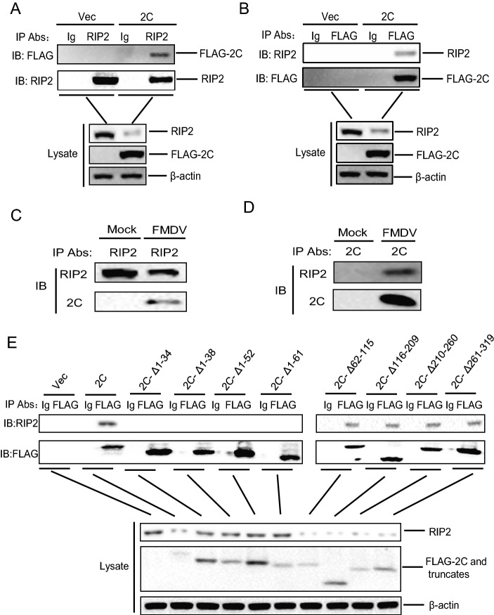
Fig. 6.
RIP2 interacted with FMDV 2C. A PK-15 cells were transfected with 10 μg FLAG–2C expressing plasmid or 10 μg empty FLAG vector. At 30 hpt, cells lysates were immunoprecipitated with anti-RIP2 antibody and subjected to western blotting. The whole-cell lysates and IP antibody-antigen complexes were analyzed by IB using anti-RIP2, anti-FLAG, or anti-β-actin antibodies. B Similar transfection in PK-15 cells and IP experiments were carried out as described above. However, the lysates were immunoprecipitated with anti-FLAG antibody. C, D PK-15 cells were mock-infected or infected with FMDV (MOI = 0.5) for 12 h. The cells lysates were immunoprecipitated with anti-RIP2 antibody (C) or anti-2C antibody (D). The antibody-antigen complexes were detected using anti-RIP2 and anti-2C antibodies. E PK-15 cells were transfected with 10 μg empty FLAG vector, 10 μg FLAG–2C expressing plasmid, or 10 μg FLAG–2C mutants expressing plasmids. At 30 hpt, cells lysates were immunoprecipitated with anti-FLAG antibody and subjected to western blotting. The whole-cell lysates and immunoprecipitated antibody-antigen complexes were analyzed by IB using anti-FLAG, anti-RIP2, or anti-β-actin antibodies.

