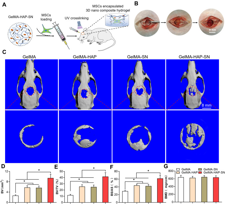
Figure 5.
Formation of the rat calvaria defect model and in vivo bone regeneration assessment. (A–B) The process of constructing an 8-mm critical-size bone defect in rats’ calvaria and the in situ UV-cross-linking of the GelMA-based MSCs embedded hydrogels. (C) Micro-CT scanning outcomes of the defective bone-healing in rats’ calvaria treated with GelMA, GelMA-HAP, GelMA-SN, and GelMA-HAP-SN hydrogel for eight weeks. The red circle indicates the initial area of the calvaria defect, which is also the area analyzed by micro-CT. (D–G) Quantification analysis of the in vivo-regenerated bone using BV, BV/TV, BS/AS, and BMD. *P < 0.05 indicates statistical difference.

