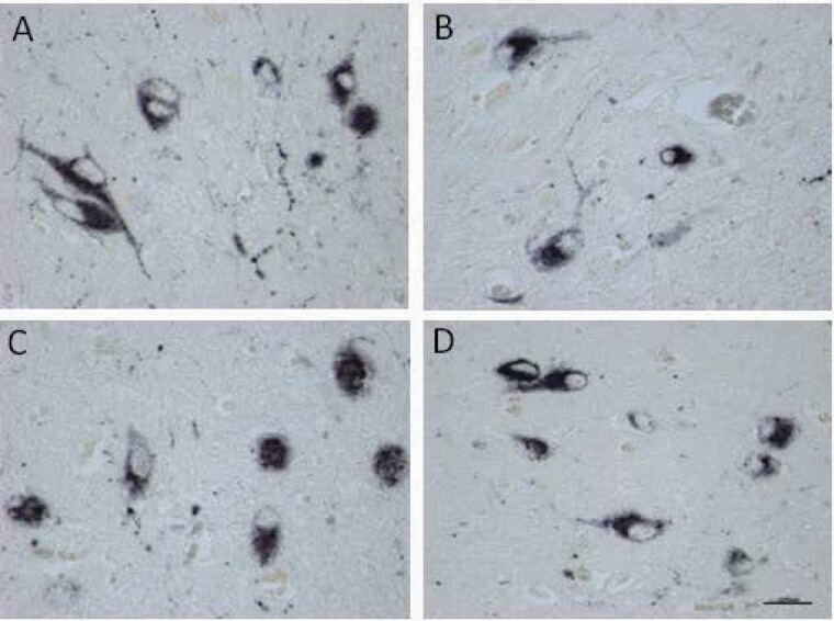
Fig. 2.
Examples of staining of hypocretin-1 cells bodies in the lateral hypothalamus of (A) a female control (NBB#2000–040). (B) a female schizophrenia patient (NBB#2010–127). (C) a male control (NBB#2010–055). (D) a male schizophrenia patient (NBB#s922-261). Bar = 0.025mm. There was no significant difference in the intensity of staining and the distribution pattern.

