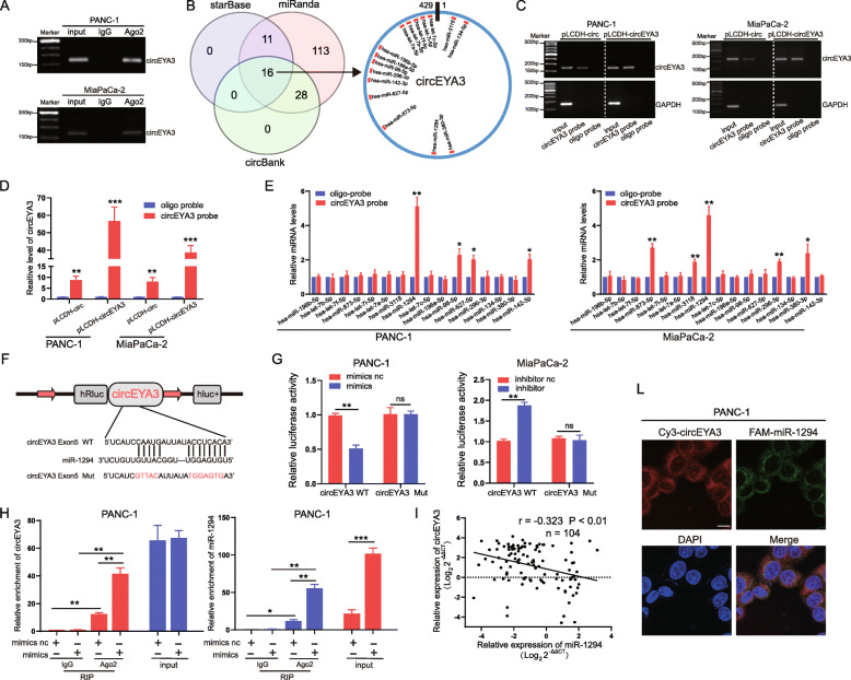
Fig. 3.
CircEYA3 acts as an efficient miR-1294 sponge in PDAC. A. The level of circEYA3 was analysed by a RIP assay in the anti-Ago2 antibody immunoprecipitate from PDAC cells. B The potential target miRNAs of circEYA3 were predicted in the circBank, starBase and miRanda databases. C and D. CircEYA3 in PANC-1 and MiaPaCa-2 cell lysates was pulled down and enriched with a specific circEYA3 probe. qRT-PCR and gel electrophoresis were used to determine the specificity and efficiency of the circEYA3 probe. E. qRT-PCR was utilized to determine the relative expression levels of 16 potential target miRNAs in precipitates from PANC-1 and MiaPaCa-2 cell lysates pulled down by the circEYA3 probe or oligo probe. F. Schematic illustration of the circEYA3-WT and circEYA3-Mut luciferase vectors. G Relative luciferase activities in PANC-1 and MiaPaCa-2 cells co-transfected with circEYA3-WT or circEYA3-Mut and the miR-1294 mimic, inhibitor or corresponding negative control. H A RIP assay was carried out with anti-Ago2 antibodies or IgG in PANC-1 cells after transfection with the miR-1294 mimic or mimic NC, and qRT-PCR was then performed to detect the enrichment of circEYA3 and miR-1294. (I) The correlation between the circEYA3 and miR-1294 expression levels in 104 paired PDAC patients was analysed by RT-qPCR and Pearson correlation analysis. L FISH assay was used to observe the cellular location of circEYA3 (red) and miR-1294 (green). Scale bar, 10 μm. *P < 0.05, **P < 0.01, ***P < 0.001; ns indicates no significance

