Fig. 5.
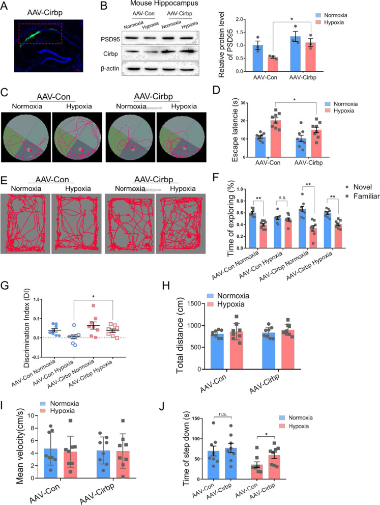
Ectopic expression of Cirbp alleviated memory dysfunction in hypobaric hypoxia exposed mice. A Fluorescence image of mouse hippocampal CA1 region (red frame area) after stereotactic injection showing Cirbp expression (green) and Nucleus (blue), scale bar = 50 μm. B Protein expression of PSD95 in mouse hippocampus following hypoxia exposure after over-expressing Cirbp (n = 3 biological replicates, two-way ANOVA, ± SEM). C, D Representative tracking plots (C) and the escape latency (D) of mice under indicated treatment in MWM test (n = 8, two-way ANOVA, ± SEM). E–I Representative locomotion tracking plots (E), exploring time on new objects (F), discriminate index (G), total distance (H) and mean velocity (I) of mice under indicated treatment in NORT test (n = 8, two-way ANOVA, ± SEM). J The latency time of step down in SIAT test (n = 8, two-way ANOVA, ± SEM). n.s. no significant, *p < 0.05, **p < 0.01

