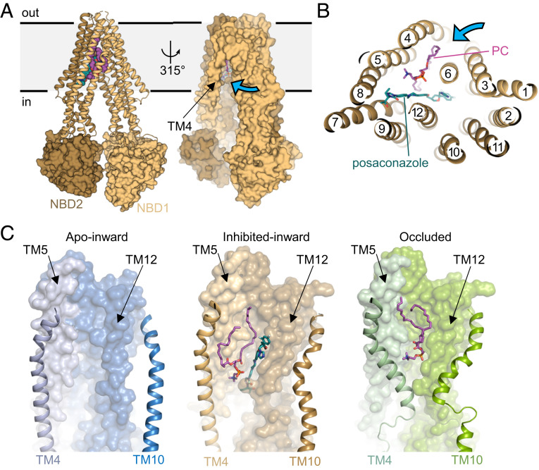
Fig. 2.
Structure of posaconazole-inhibited ABCB4. (A, Left) TMDs are shown as ribbons and NBDs as surfaces. The N- and carboxyl-terminal halves of ABCB4 are colored differently. Posaconazole and PC are shown in sphere representation and colored green and purple, respectively. (Right) Surface representation. The blue arrow shows the side opening to the membrane. (B) Horizontal slice through a ribbon representation, with posaconazole and PC shown as green and purple sticks. (C) Close-up views of the TMDs in distinct states. Bound PC and posaconazole are shown in sticks and colored as in B. The TM helices 4 and 10 undergoing conformational changes are shown as ribbons. Posaconazole prevents bound PC from reaching the central pocket and TM4 from adopting a kinked conformation.

