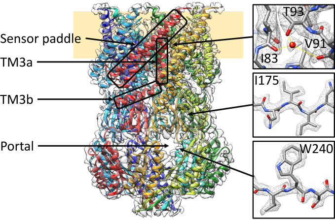
Fig. 1.
Structure of open MscS solubilized in LMNG. MscS consists of seven identical subunits, shown in different colors, and is organized in a membrane domain (Top) and a cytosolic domain (Bottom). In the membrane domain, TM1 and TM2 form the sensor paddle, TM3a the pore, and TM3b connects the two domains. The cytosolic domain shapes a vestibule that can be accessed through portals. Some close-ups of the densities are shown on the right together with the atonic model (oxygen: red, nitrogen: blue, carbon: gray). Yellow lines indicate the coordination of the water. The sample was obtained under condition 2b (see Results).

