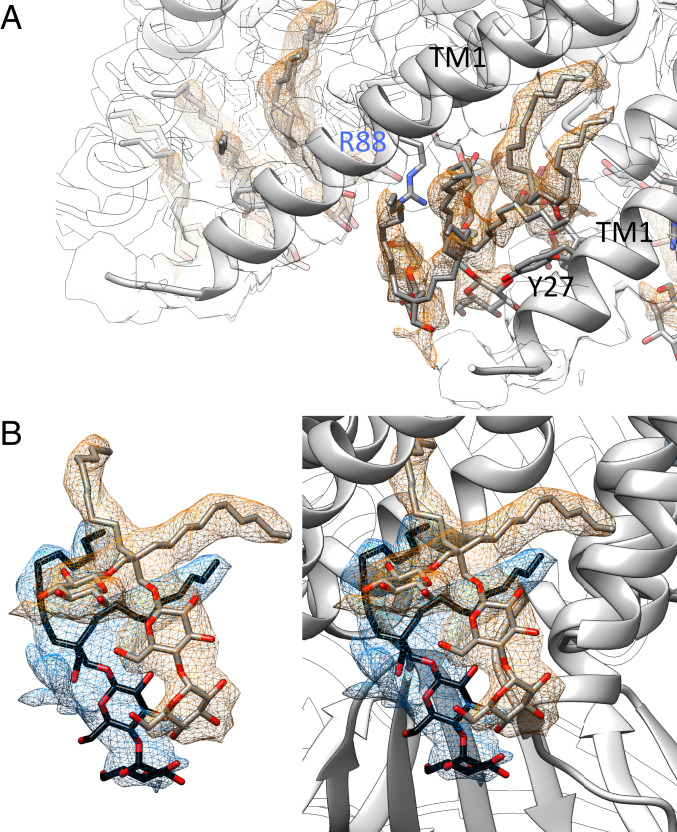
Fig. 5.
Stacking of LMNG molecules. (A) On the periplasmic side, three LMNG molecules stack on each other in between the TM1 helices. The best-resolved one is at the loop between TM2 and TM3a (see Fig. 4D). The middle one is clamped by R88 and Y27. The headgroups face toward the water-filled funnel in the center, while the hydrophobic tails face outwards into the micelle. The protein density is shown as an outline at the same threshold. (B) Two LMNG molecules stack on the cytosolic side covering a hydrophobic opening. The image on the right side also shows the context of the protein structure.

