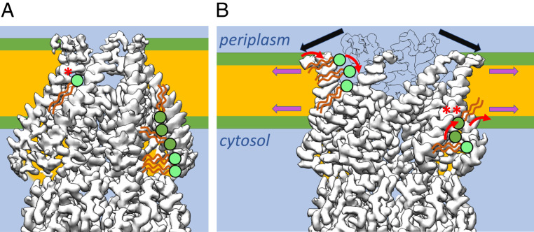
Fig. 6.
Scheme of lipid interaction with MscS during gating. (A) MscS in the closed and (B) open conformation schematically together with the membrane and interacting lipids. For clarity, only on the left side of MscS examples for interacting lipids from the periplasmic leaflet are shown (for each conformation). On the right side of MscS interacting lipids from the cytosolic leaflet are exemplified. Lipids that were directly seen in the structures or were suggested by the positions of LMNG are shown in light green. (A) In the closed conformation, a lipid is locked between two adjacent sensor paddles in the middle of the membrane with its headgroup facing the funnel (*). There is no pathway for its charged headgroup to cross to the cytosolic leaflet, and the pathway to the periplasmic leaflet is blocked by TM1 helices. On the cytosolic side, large hydrophobic pockets are filled with lipids from the cytosolic leaflet. (B) Additional tension in the membrane leads to thinning of the membrane and is highest at the onset of the hydrophobic chains (magenta arrows). Our model proposes that lipids in the pockets in this area (**) will initiate the conformational change in MscS. The N-terminal domains at the periplasmic interface are not completely resolved, but this region of MscS shows a substantial outward movement (black arrows; an outline of the closed conformation is shown for comparison), which leads to the separation of the TM1 helices. Now, the bulk water phase in the funnel would be exposed to the membrane core. To prevent that from happening, lipid head groups must move from the periplasmic leaflet into these gaps and equilibrate with the lipid trapped in the closed state. The amphipathic N-terminal domains may also sense tension directly in the membrane in the periplasmic leaflet.

