Fig. 2.
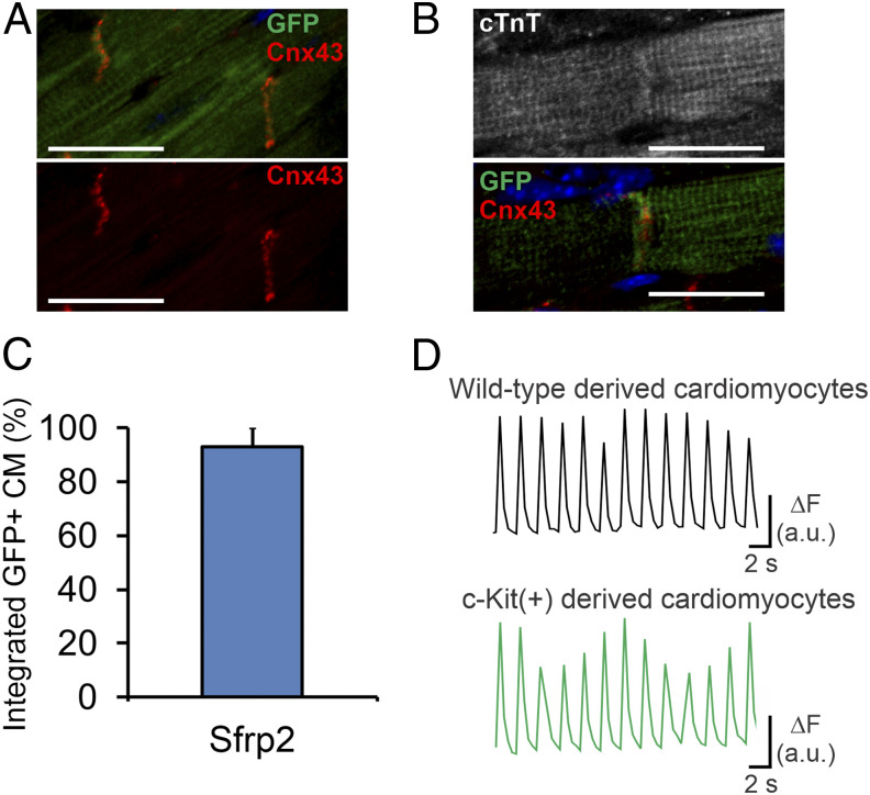
Cardiomyocytes generated via Sfrp2 are physiologically normal and integrate with the myocardium. Following tamoxifen administration to activate the Cre, cKitCreERT2/mTeG mice were subjected to MI. Two days after injury, mice received either vehicle or Sfrp2. Two months after injury, cardiac tissue was analyzed. Sfrp2-derived (eGFP+) cardiomyocytes formed gap junctions with (A) adjacent Sfrp2-derived (eGFP+) cardiomyocytes as well as (B) preexisting (eGFP−) cardiomyocytes, as shown by connexin-43 staining. (Scale bar, 50 μm in A or 25 μm in B.) (C) Quantification of the percentage of Sfrp2-derived (eGFP+) cardiomyocytes, which show apparent integration. n = 7 individual animals. (D) EC coupling in eGFP− and eGFP+ cardiomyocytes. Representative examples of calcium transients obtained from Fura-2–loaded wild-type (eGFP−) and Sfrp2-derived (GFP+) cardiomyocytes during pacing at 0.5 Hz with electric field stimulation.

