Fig. 4.
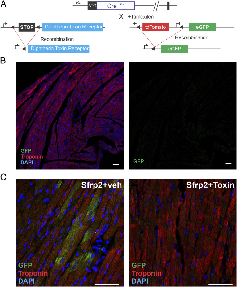
Genetic ablation prevents Sfrp2-induced cardiomyogenesis. (A) cKitCreERT2/mTmG mice were crossed with a DTR strain to selectively ablate cKit(+) cells in the heart. Mice (cKitCreERT2/mTmG/DTR) were injected with tamoxifen (0.5 mg/mouse) for 14 consecutive days. In the final 7 d of tamoxifen treatment, dipththeria toxin (100 ng/mouse) was injected daily to ablate cKit(+) positive cells. Four days later, treatment mice were subjected to MI. Two days later, mice were injected with Sfrp2 (0.5 µg) or vehicle at the infarct border zone. (B) cKitCreERT2/mTmG/DTR mice were injected with tamoxifen (0.5 mg/mouse) for 14 consecutive days. In the final 7 d of tamoxifen treatment, dipththeria toxin (100 ng/mouse) was injected daily to ablate cKit positive cells. Heart tissue was analyzed for cardiac troponin-T and eGFP expression by confocal microscopy. n = 3. (C) Representative confocal images of 2 mo post-MI hearts. Colocalization of eGFP and the cardiac marker cardiac troponin-T was determined by confocal microscopy. (Scale bar, 50 μm.) n = 5 per group.

