Fig. 5.
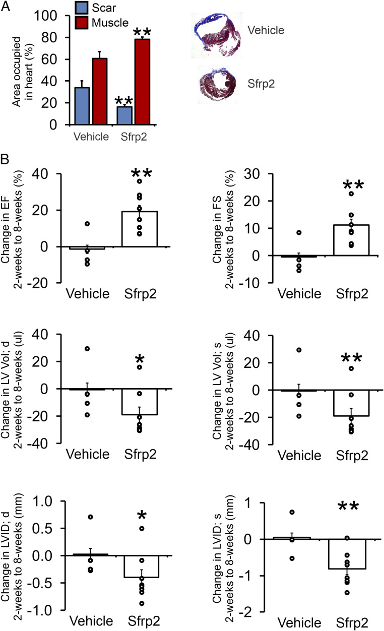
Sfrp2-induced cardiomyogenesis is associated with improved therapeutic outcomes. Mice (cKitCreERT2/mTmG) were injected with tamoxifen (0.5 mg/mouse) for 14 consecutive days. Four days after tamoxifen treatment, mice were subjected to MI. Two days after injury, mice were injected with Sfrp2 (0.5 µg) or PBS at the infarct border zone. (A) Analysis of Masson’s trichrome staining for cardiac muscle (red) and fibrosis (blue). Sections were taken at 0.5 mm and 1 mm below the Sfrp2/vehicle injection point. n = 4 (vehicle) or 8 (Sfrp2); t test, **P < 0.01. Representative Masson’s trichrome–stained cardiac sections are shown. (B) Cardiac function was assessed by echocardiography immediately prior to injury, 2 wk after injury, and finally 8 wk after injury. Raw cardiac function data are provided in SI Appendix, Table S1. Comparisons were made between 2 and 8 wk postinjury in the control and Sfrp2 groups. The graphs show the comparison for each animal (open circle) as well as the average value in each group (bar). n = 4 (vehicle) or 8 (Sfrp2). t tests were carried out between the control and Sfrp2 groups; *P < 0.05, **P < 0.01.

