Fig. 1.
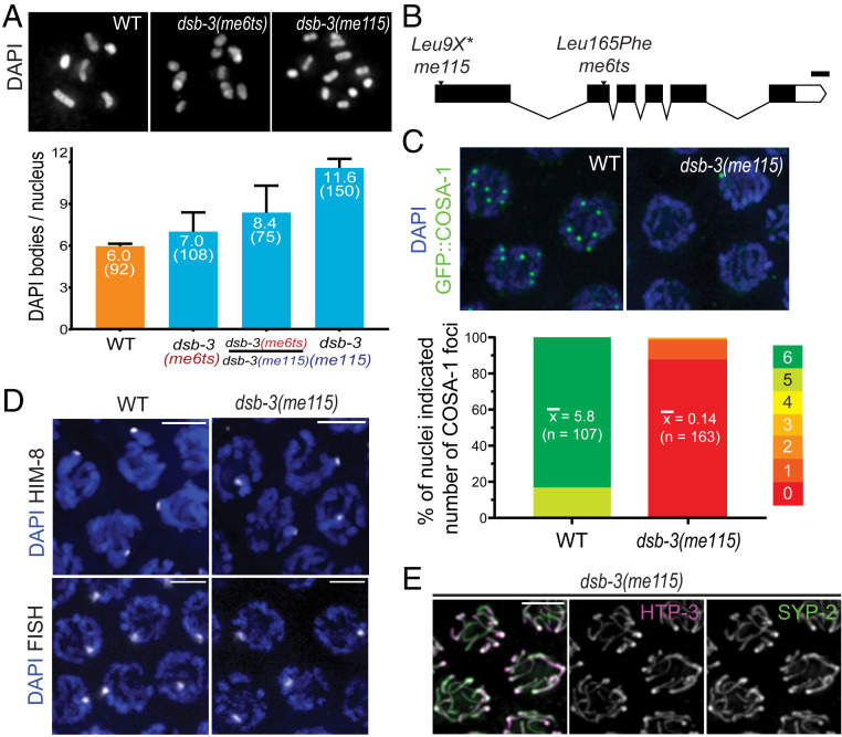
Identification of dsb-3 as a gene required for meiotic CO formation. (A, Top) Representative images of diakinesis-stage oocyte nuclei from adult worms fixed at 1 d after L4. (Left) Wild-type (WT) nucleus with six DAPI bodies corresponding to six pairs of homologs connected by chiasmata (bivalents). (Middle) dsb-3(me6ts) nucleus with nine DAPI bodies (three bivalents and six univalents). (Right) dsb-3(me115) nucleus with 12 DAPI bodies (all univalents). (A, Bottom) Quantification of DAPI bodies/ nucleus; error bars indicate standard deviation, and numbers in parentheses indicate the numbers of nuclei assayed. For all pairwise comparisons, Mann–Whitney P values were <0.0001. Assays for WT and dsb-3(me115) homozygotes were performed at 20 °C; assays for dsb-3(me6ts) homozygotes and dsb-3(me6ts)/dsb-3(me115) heterozygotes were performed at 25 °C. (B) Schematic showing the dsb-3 gene structure, with the positions and nature of mutations used in this work; white boxes represent untranslated region sequences, black boxes represent exons, and lines indicate introns. (Scale bar: 100 bp.) (C, Top) Immunofluorescence images of GFP::COSA-1 foci, which correspond to the single CO site on each homolog pair in late pachytene nuclei. WT nuclei have six foci, while foci are reduced or absent in the dsb-3(me115) mutant. (C, Bottom) Stacked bar graphs showing the distribution of GFP::COSA-1 foci counts in nuclei from WT and dsb-3(me115) mutants. Mean numbers of foci per nucleus are indicated, with the numbers of nuclei assayed in parentheses; Mann–Whitney P < 0.0001. (D) Homolog pairing assayed by immunofluorescence of X-chromosome pairing center binding protein HIM-8 (Top) or FISH detecting a 1-Mbp segment of chromosome II (Bottom) in pachytene nuclei of whole-mount gonads. A single focus is observed in each nucleus, indicating successful pairing. (Scale bars: 3.2 µm.) (E) Immunofluorescence image of SC components in late pachytene nuclei in a whole-mount gonad from the dsb-3(me115) mutant. Axis protein HTP-3 and SC central region protein SYP-2 colocalize in continuous stretches between chromosome pairs, indicating successful synapsis. (Scale bar: 3.2 µm.)

