Fig. 2.
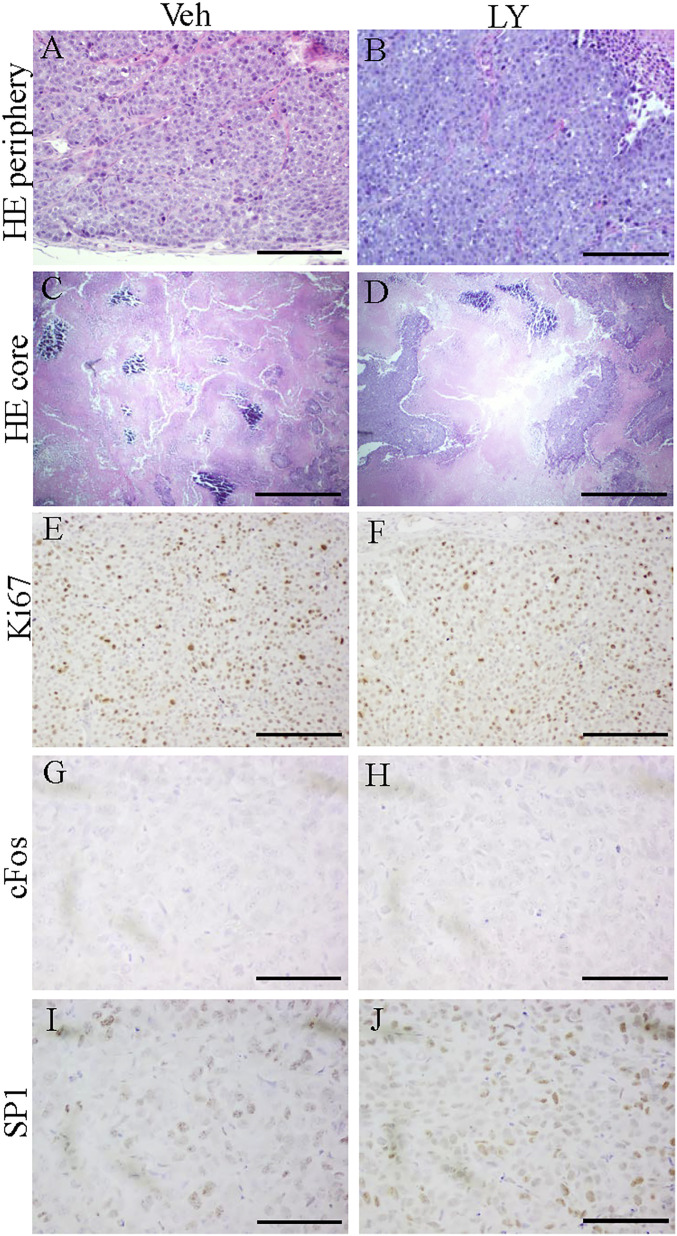
H&E staining and expression of Ki67, cFOS, and SP1 in TNBC PDXs. H&E staining of five PDXs in each treatment group demonstrated that cancer cells were densely packed together in peripheral zones (A and B) and that there were large necrotic regions in the core of the PDX tumors (C and D). Ki67 was highly expressed in both the Veh-treated group and LY500307-treated group (E and F). Expression of cFOS was not detected by immunohistochemistry staining (G and H). SP1 was highly expressed in Veh-treated or LY500307-treated PDX tumors (I and J). (Scale bars, 100 μm [A, B, and E–J] and 500 μm [C and D].)

