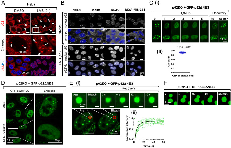
Fig. 1.
Nuclear p62 condensates are formed via LLPS. (A) DMSO (Left) or LMB-treated (Right) HeLa cells were stained with a p62 antibody and Hoechst (Ho) and imaged using 2D confocal microscopy. Arrows point to nuclear p62 foci. (B) The indicated cells were treated with either DMSO (Upper two lines) or LMB (Lower two lines), and stained with anti-p62 and Ho. For highlighting nuclear p62, the cytosolic p62 was hidden by the software when only p62 was stained. (C) Time-lapse imaging of GFP-p62∆NES condensates disruption and recovery following 1,6-HD treatment. (D) Live cell images of the indicated cell treated with the E1 inhibitor MLN-7243. (E) (i) FRAP (Lower, red line surrounded circles) of p62 foci. Time-lapse images (Upper) were taken from bleached cells; (ii) quantification of fluorescence taken from 18 p62 condensates. (F) Time-lapse images of fusion of p62 condensates.

