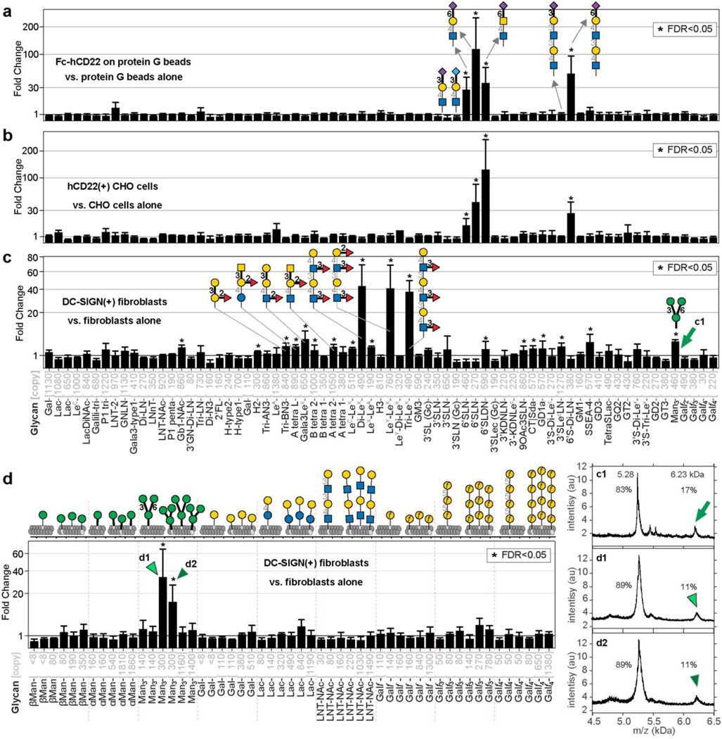
Fig. 5: LiGAs measure affinity and avidity-based responses in cell-displayed GBPs.
a, Binding of LiGA-71 to construct hCD22-Fc normalized by binding of the same LiGA to protein-G beads, n=3. b, Binding of LiGA-71 to hCD22(+) CHO cells normalized by binding of the same LiGA to CHO cells, n=4. c, Binding of LiGA-71 to DC-SIGN(+) fibroblasts normalized by binding of the same LiGA to fibroblasts, n=4. d, Binding of LiGA 9×6 to DC-SIGN(+) fibroblasts performed analogously to (c), n=3. Phage virions that display 300 copies (d) or 460 copies (c) of core trimannoside exhibit significant binding to DC-SIGN clusters on the cell surface, virions with <150 copies or >1200 copies exhibit insignificant enrichment. Calculations of FC, FDR and error bars are identical to those described in Fig. 2h, Fig. 3a, and 4b–d.

