Figure 2.
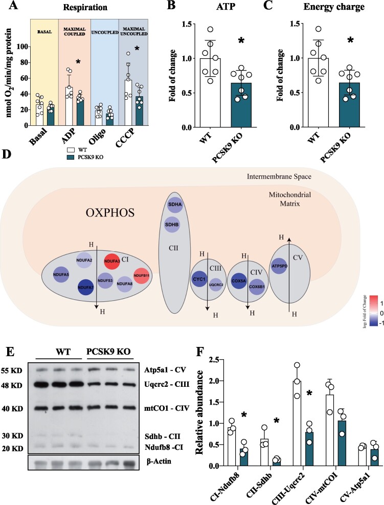
Pcsk9 deficiency is associated with mitochondrial dysfunction. (A) The oxygen consumption rate was investigated in the heart of Pcsk9 KO mice and oxygen consumption was measured at basal, maximal coupled, uncoupled and maximal uncoupled conditions (Basal, P = 0.39; ADP, P = 0.04; Oligo, P = 0.24; CCCP, P = 0.04). Data are shown as mean ± SD; n = 7 mice per group. (B and C) Adenosine triphosphate (P = 0.01) quantification and energy charge (P = 0.01) of the heart. Data are shown as fold of change ± SD; n = 7 mice per group. (D) Relevant proteins of ETC are displayed. (E) Representative image of western blot of ETC complexes on heart lysate is showed. (F) Proteins quantification is normalized to beta-actin expression (CI, P = 0.01; CII, P = 0.02; CIII, P = 0.008; CIV, P = 0.08, CV, P = 0.59). Data are shown as mean ± SD; n = 3 mice per group. Non-parametric t-test was used to compare each group (*P < 0.05).

