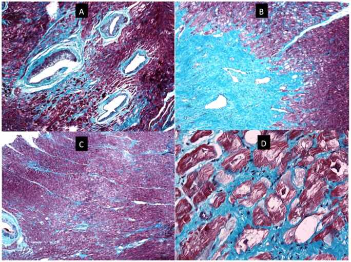
Figure 7.
Histopathology images for index patient # 23. (A) A section of myocardium stained with Masson trichrome. There are four dysplastic vessels in the field. The muscle of the tunica media is irregularly distributed around the circumference of the vessels and in places is almost absent. There is accompanying mural fibrosis. The surrounding myocardium shows patchy interstitial fibrosis and focal myocyte vacuolation. (B) Low power view of myocardium showing a collagenous scar. The scar tissue contains thin-walled ectatic vessels and interdigitates with the surrounding myocardium that is vacuolated and shows foci of fine interstitial fibrosis (Masson-trichrome stain). (C) Low-power view of myocardium stained with Masson trichrome. The field shows an area of central pallor caused by a localized focus of vacuolated myocytes. There is fine interstitial fibrosis. There is no myocyte disarray. A dysplastic vessel is visible at the left edge of the field. (D) High-power view of a section of myocardium stained with Masson trichrome. It contains myocytes with irregular central areas of clearing of the cytoplasm to give vacuoles. Some of the vacuoles are traversed by fine strands of cytoplasm and other contain abundant normal mitochondria (seen as small red dots). Many of the vacuoles, however, are empty. Periodic acid-Schiff staining was negative. Desmin and plakoglobin staining was normal (Supplementary material online).

